

In the U.S. today, more than 100,000 people are waiting for an organ transplant and 130 million Americans will visit the emergency room this year. In the not-so-distant future, doctors could use machines that directly repair tissues by depositing new layers of muscle or skin, even creating and installing new organs.
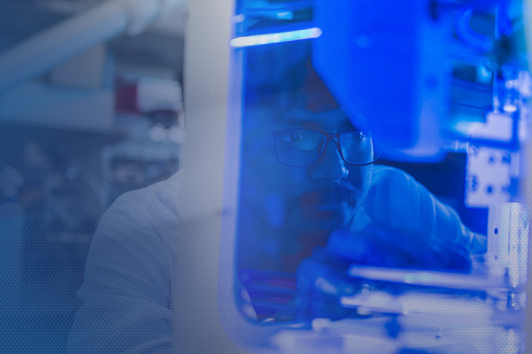
This scenario is not science fiction for Penn State researcher Ibrahim T. Ozbolat, an associate professor of engineering science and mechanics, biomedical engineering, and neurosurgery. Ozbolat is using 3D bioprinting to create a range of materials for potential use in human health, including printable bone, skin, and tumor cell models.
At Penn State, researchers are working directly with industry to use this technology in novel ways to address pressing problems in human health and other areas. The ability of Penn State’s researchers to collaborate with colleagues across disciplines to address every component of a problem and its solution is a key advantage to the University’s cutting-edge approach to additive manufacturing.
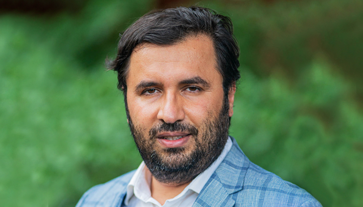
Ibrahim Ozbolat, Hartz Family Career Development Professor of Engineering Science and Mechanics and Biomedical Engineering
WATCH
Bioprinting for You
“In the future, bioprinting will play a critical role in every aspect of the healthcare system. It could directly enable the creation of transplantable organs. It will reduce the need for organ donations. One day, this technology will enable us to better understand diseases and reduce the need for animal testing.”
Printing with Biomaterials
“I can envision a time when a patient might lie under a bioprinter and have new skin printed directly onto a wound,” Ozbolat says. Already, Ozbolat and his lab group have reported success in printing both bone and soft tissue onto the skulls of rats.
“Repairing injuries to the skin and bones of the skull is particularly difficult given the many layers of different types of tissues involved,” he says. “Trying to work with these two materials at one time is an even greater challenge.”
Currently, Ozbolat explains, fixing something as complex as skull injuries currently requires the use of skin and bone from another part of the patient’s body. This can require additional surgery, or sourced materials from a cadaver, which runs the risk of rejection by the patient’s immune system. For their study, Ozbolat and his colleagues instead created a printable bone material using a mixture of collagen; chitosan, sugar from the outer skeleton of shellfish; nano-hydroxyapatite, a component of tooth enamel; and bone morphogenetic protein-2, an FDA approved growth factor for bone regeneration. For the skin, they used collagen and fibrinogen, a protein made in the liver that aids in blood clotting.
After precisely scanning a rat’s skull defect, Ozbolat explains, the 3D printer followed the 3D “blueprint,” extruding the bone material onto the wound, followed by a barrier material, and then the skin material. The whole process took less than five minutes. After separately repairing the 6-millimeter-wide hole in the skin and the 5-millimeter-wide hole in the bone, they moved on to repair both during the same surgical procedure. “There is no surgical method for repairing soft and hard tissue at once,” notes Ozbolat.
The next step, he says, is to add compounds that can help to facilitate vascularization, since blood flow to a bone is especially important for healing. He and his team are already working with neurosurgeons, craniomaxillofacial surgeons, and plastic surgeons at Penn State Hershey Medical Center to translate this research to human applications.
In addition to repairing skin and bone, Ozbolat and his team are using 3D bioprinting to help in the study of breast cancer. In a recent study, the team generated tumor models, called tumor spheroids, to study how a tumor cell’s distance from nearby endothelial cells—cells that line the walls of blood vessels—and fibroblasts—connective tissue cells—influences its ability to grow. The closer a tumor cell is to an endothelial cell or fibroblast, they found, the more aggressively it is likely to spread.
“In research like this, it is important to maintain precision in the variables that are being tested,” says Madhuri Dey, a Ph.D. candidate in chemistry. “In this project, 3D printing allows us to precisely adjust the position of the tumor with respect to the main blood vessel so we can observe the effects of distance on tumor growth. Using a natural tumor would introduce too much variability.”
“In this project, 3D printing allows us to precisely adjust the position of the tumor with respect to the main blood vessel so we can observe the effects of distance on tumor growth.” —Madhuri Dey, Ph.D. candidate in chemistry
Additive Manufacturing
The term additive manufacturing describes the use of 3D printing to make functional components in a manufacturing setting. The process is “additive” because it produces an object by building it up one layer at a time. By contrast, subtractive manufacturing creates components by removing material until the final part is complete. The additive process, by its nature, is both more flexible and far less wasteful.
The process begins with a 3D model: a computer-aided design (CAD) representation of the object that specifies precisely how much material—plastic, metal, clay, or biomaterial—needs to be deposited, and where. In traditional manufacturing, a 3D printer reads its instructions, extrudes a plastic filament from a nozzle onto the print bed in a specified manner or a laser melts metallic powder layer by layer to form a part. After completing the base layer, the printer adds additional layers until the item is complete.
In a way, researchers believe that they can print almost anything they can imagine, allowing us to produce traditional products more affordably and sustainably and to create entirely new products that may transform the way we travel, build homes, and manage our health.